|
Lymphatic Obstruction Lymphatic obstruction is caused by Wuchereria bancrofti. Wuchereria bancrofti can be detected from thick smear blood examination from blood sample taken at 10 pm to 4 am due to Wuchereria microfilariae transmission into the blood at night. Wuchereria bancrofti is a nematode roundworm. Culex mosquito or Anopheles mosquito are considered as vector in transmitting Wuchereria bancrofti. The larvae will penetrate the skin after bite from the mosquito. The larvae will migrate to the regional lymph nodes. Larvae may develop into adults after 6 months to 12 months incubation period. There will be production of microfilariae larvae which will migrate into the blood at night time. As a consequences, there will be obstruction and edematous lymphatic lesion. Patient may present with symptoms such as chills, lymphadenitis, eosinophilia and fever. Chronic cases may lead to inflammation and obstruction of the lymphatic system. This will finally results in elephantiasis or filariasis. The prevention methods may include considering insect repellant or mosquito nets. Diethylcarbamazines is given to the patient to kill the worms and surgery is considered to relieve the obstruction.  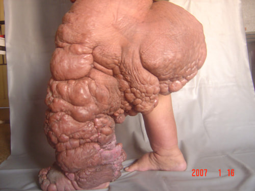
0 Comments
Pinworm infection Pinworm is also known as Enterobius vermicularis. Enterobius vermicularis is also known as nematode roundworm. The common signs and symptoms of infection caused by pinworm may also include itching of the perianal region which later lead to scratching of the intestine. The complication of pinworm infection may include vaginitis due to migration of Enterobius vermicularis into the vagina. The mode of transmission of pinworm infection is via ingestion of eggs from fecal oral spread. The eggs will hatch in the small intestine while the larvae migrate to the colon and grow into adults. The female adults may later lay eggs in the perianal region. Perianal sampling is taken by using adhesive tape and an applicator to detect the present of the egg. The prevention steps taken to stop pinworm infection may include good personal hygiene. The treatment may include mebendazole and pyrantel pamoate which kill the pinworm. Sporotrichosis Sporotrichosis is caused by fungus Sporothrix schenckii. The common symptoms and signs of sporotrichosis may include the formation of ulcers and nodules along the draining of lymphatic chain and at the sites of inoculation. Sporotrichosis is mostly seen in gardeners. Sporotrichosis is transmitted from soil, decaying vegetation or from puncture wounds. Sporonthrix schenckii is a dimorphic fungus. The common sources of infection are baled hay and rose thorns. Sporotrichosis will induce an inflammatory responses. Sporonthrix schenckii will inhibit the action of neutrophils by producing melanin. The prevention steps taken to avoid sporotrichosis may include wearing protective clothing while handling hay bales or roses thorn ( gardener) and wearing protective gloves.Patient with sporotrichosis is treated with amphotericin B, itraconazole and potassium iodide. Pneumocytosis Pneumocytosis is also known as pneumocystis pneumonia which is caused by pneumocystis jiroveci. The laboratory investigation needed to confirm the morphological structure of pneumocystis jiroveci may include Gomori methenamine silver stain of the sputum and bronchoalveolar lavage Pneumocystis pneumonia is characterized by dyspnea, cough and fever. Pneumocystis pneumonia is commonly seen in HIV- AIDS patient as well as malnourished children and premature infant. The common mode of transmission of pneumocystis pneumonia is respiratory droplets. Pneumocystis jiroveci will block the gaseous exchange process in the lung by accumulating in the lumen of the airway via attaching to the pneumocyte of the alveolar and inducing the infiltration of the inflammatory cells. Pneumocystis pneumonia may spread into the liver, bone marrow, spleen and lymph nodes.The common cause of death in HIV- AIDS patient is related to pneumocystosis / pneumocystis pneumonia. The treatment / prophylaxis of pneumocytosis / pneumocystis pneumonia may include trimethoprim and sulfamethoxazole. Lobomycosis Lobomycosis is caused by loboa loboi . Lobomycosis may lead to formation of keloid ( hard painless nodules) on the ear, face and upper extremities. Lobomycosis may also occur ( loboa loboi infection) in dolphins. Lobomycosis occur due to traumatic inoculation. Loboa loboi is detected by the present of yeast cells in long chain which can be identify by direct microscopic of excised tissue in potassium hydroxide preparation. The treatment of lobomycosis may include surgical excision of the lesions. Infectious Mononucleosis Infectious mononucleosis is caused by Epstein Barr virus. The mode of transmission of Epstein Barr virus is due to saliva. Infectious mononucleosis may present with infection of the oropharygeal cells which later spread to B lymphocytes which lead to latent infection. Epstein Barr virus may also lead to mitogenesis of the B cells with immortalization of the B cells. Epstein Barr virus may lead to Epstein Barr virus associated tumor ( the present of Epstein Barr virus DNA in tumor) due to immortalization of B cells. The common symptoms and signs of infectious mononucleosis may include fatigue, headache, malaise, pharyngitis, fever, splenomegaly, raised liver enzyme and lymphadenopathy. The common complication of infectious mononucleosis may include nasopharyngeal carcinoma, Burkitt lymphoma and Hodgkin lymphoma as well as lymphoproliferative disorders. Diagnosis test such as Monospot test is important in diagnosing infectious mononucleosis related Epstein Barr virus. In this case, the heterophile antibody positive result ( positive monospot test) may confirm the disorder. Other test may include the identification of the Downey cells / atypical lymphocytes in the blood smear and Epstein Barr virus serological test. Rhinosporidiosis Rhinosporidiosis may present with painless pedunculated nasal polyps. Rhinosporidiosis may be caused by Rhinosporidium seeberi. Rhinosporidiosis mostly happen due to traumatic inoculation in divers. Rhinosporidiosis may be reveal under direct microscopic examination of nasal discharge or excised tissue in potassium hydroxide solution as endospores and spherules. Rhinosporidiosis is associated with fish as the natural reservoir. There will be formation of abscess with infiltration of inflammatory cell and necrosis of the tissue. The common treatment of rhinosporidiosis may include amphotericin B ( injection) or surgical removal of the lesions. Hookworm infection There are two species of hookworm which may include ancylostoma duodenale and necator americanus. Hookworm infection is diagnosed by the present of egg in the stool. Hookworm infection may present with symptoms and signs such as allergic rash, pneumonitis ( due to larval lung migration) loss of weight, fatigue and microcyctic hypochromic anemia ( adult hookworm feed on the blood, patient may loss 0.2 ml blood per day). The eggs of the hookworm from human feces may present in the soil which later develop into larvae in the soil. Larvae may penetrate into the skin which lead to infection. Larvae will transfer to the lung and finally end up in the small intestine as adult worms. Adult worms may produce more than 10 000 eggs which will be passed in the stool. Hookworm infection can be prevented by proper disposal of human waste and wearing the shoes. Hookworm can be killed with mebendazole and pyrantel pamoate. Subacute Sclerosing Panencephalitis Subacute sclerosing panencephalitis is caused by mutant measles virus. Patient with subacute sclerosing panencephalitis may be detected by high measles virus antibody levels in cerebrospinal fluid and serum. Subacute sclerosing panencephalitis may present with inflammation and demyelination. Subacute sclerosing panencephalitis is caused by variant of measles virus with mutation in the matrix protein which important in assembly and budding of the virus. There will be no antibody to matrix protein. Patient with subacute sclerosing panencephalitis may present with changes in the intellectual and personality with period of muscular spams ( myoclonic jerk) , blindness, spasticity and death. Measles virus is transmitted mostly via respiratory droplets. Measles virus may escape immune reaction due to action of fusion protein ( cell to cell fusion). Subacute sclerosing panencephalitis may occur in children 6 years after acute attack of measles and presented with late progressive neurological disorders. Patient is given live attenuated measles virus vaccine to prevent this condition. Astrovirus infection Astrovirus infection may lead to acute gastroenteritis. Astrovirus infection typically affecting children and infant. Astrovirus will damage the enterocytes by infecting the epithelial cells of the intestine. Patient may present with symptoms and signs of acute gastroenteritis such as fever, abdominal pain and watery diarrhea. The common mode of transmission of astrovirus is via fecal oral route. Astrovirus can be detected via polymerase chain reaction or viral antigen assay. |
Kembara's Health SolutionsDiscovering the world of health and medicine. Archives
June 2023
Categories
All
|
 RSS Feed